DEXIS CariVu™ — Caries (Cavity) Detection Device
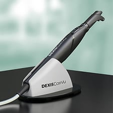 A brilliant new approach to cavity (caries) detection. By hugging the tooth and illuminating it in safe, near-infrared light, CariVu’s transillumination technology makes the enamel appear transparent while porous lesions or cracks trap and absorb the light. This allows the doctor to see through the tooth, exposing its structure and the actual structure of any carious lesions with very high accuracy. The CariVu technology allows us to get another perspective on suspicious areas that may show up on x-rays, which means we can do a better job than ever at detecting decay in its early stages.
Cavitron JET Plus Ultrasonic Scaler and Air Polishing System
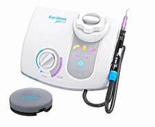 A Cavitron Ultrasonic Scaler is a dental hygiene tool that uses high frequency sound waves to clean teeth. The Cavitron JET Plus used at Ufberg Dental is a state of the art instrument that has proven to be a very successful tool in removing tartar from both above and below the gumline. Instead of scaling the teeth with bulky hand scalers, the Cavitron Ultrasonic Scaler uses oscillating sound waves to gently vibrate the tartar away from your teeth. The Cavitron unit is so gentle that it can even be used for deep cleaning and gum therapy without the need for anesthesia. Having your teeth cleaned with a Cavitron is a safe effective and painless alternative to traditional cleaning methods.
Some of the advantages of the Cavitron Ultrasonic Scaler include:
- Faster than traditional teeth cleaning methods
- A pleasant alternative to hand scaling
- Safe and effective method to clean teeth
Digital X-Rays
 Digital radiography is a form of X-ray imaging, where digital X-ray sensors are used instead of traditional photographic film. Advantages include time efficiency through bypassing chemical processing and the ability to digitally transfer and enhance images. Also less radiation can be used to produce an image of similar contrast to conventional radiography.
Instead of X-ray film, digital radiography uses a digital image capture device. This gives advantages of immediate image preview and availability; elimination of costly film processing steps; a wider dynamic range, which makes it more forgiving for over- and under-exposure; as well as the ability to apply special image processing techniques that enhance overall display of the image.
Digital X-rays can be sent to a computer to be recorded and saved.
Oral ID
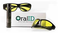 OralID’s fluorescence technology uses a blue light that allows a clinician to identify oral cancer, pre-cancer and other abnormal lesions at an earlier stage, thus saving lives.
ARESTIN®
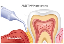 ARESTIN® is a prescription antibiotic approved by the Food and Drug Administration (FDA). It is used together with scaling and root planing (SRP) and is placed by your dentist for the treatment of periodontal (gum) disease.
If you have periodontal (gum) disease, SRP is needed to help improve the health of your teeth and gums. Bacteria are the cause of gum disease. That’s why your dental professional may recommend ARESTIN®. It is an antibiotic that helps kill bacteria at the root of the problem. Take your dental professional’s advice—choose ARESTIN®.
ARESTIN® contains microspheres—tiny particles—that are smaller than grains of sand and are not visible to the eye. The microspheres are filled with the antibiotic minocycline hydrochloride. These microspheres release the antibiotic over time, killing bacteria so your gums can heal better than with SRP alone.
Intra-Oral Camera
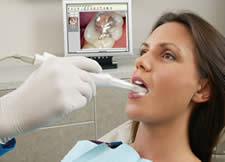 In a nutshell, an intraoral camera is a small video camera that takes an X-ray of the outside of the gum or tooth. The intraoral camera resembles an oversized pen and although usage varies depending on the model-type, this image-taking device is typically outfitted with a disposable protective sheath for each new patient. While simultaneously viewing a monitor, the dentist inserts the camera into a patient’s mouth and gently shifts it about so that images can be taken from a variety of angles.
The intraoral camera is especially useful during dental restoration procedures. For example, if you were to have an amalgam tooth filling replaced with a composite resin filling, your dentist could use the intraoral camera to take “before and after” pictures and display the results simultaneously for you to see!
In addition to being a great diagnostic tool, the intraoral camera is a fantastic educational aid. Instead of merely explaining to you what’s happening inside your mouth, your dentist can actually show you. And, unlike conventional X-ray images that require processing time, there is no development time associated with intraoral cameras: The immediately available images that this tool renders can be a great time-saver for both you and your dentist.
Soft Tissue Laser
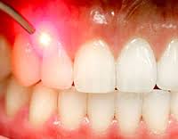 Laser energy is a form of light that is highly focused and monochromatic, or one color. A monochromatic color allows laser light to be set at a certain wavelength to attract certain molecules.
In laser dentistry, these molecules are hemoglobin, water, and melanin – the molecules that make up your gum tissue.
Because the laser is only attracted to your gum tissue, it’s a very safe tool to use. It will not affect hard tooth structure or any metal you may have in your mouth. It only affects the area of tissue your dentist wants to treat.
A laser is more precise, causes less pain, and prevents bleeding better than traditional tools used on soft tissues. The highly focused laser light cauterizes nerve endings, coagulates blood vessels, sterilizes the surgical site, and increases the speed of healing. Instantly cauterizing nerve endings greatly reduces pain during the procedure and after.